Carpal Joint Orthoses
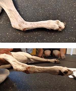
Injury to the carpus is common and can be complex because the joint itself is complex. The carpus as a biomechanical structure must be thought of as 3 joints, 7 carpal bones, 2 antebrachial bones, and 4 or 5 metacarpal bones. There are multiple ligaments holding this structure together, collectively referred to as the collateral ligaments and the palmar fibrocartilage. Lastly, primary muscles of the carpus and digits including the flexor carpi ulnaris, flexor carpi radialis, and superficial and deep digital flexors play a significant role in distal limb function. Because of the role of the digital flexors, the digits themselves must be considered in carpal injuries. Primary carpal injuries can occur at any of the 3 joints (antebrachiocarpal, middle carpal, or carpometacarpal); additionally, any of these bones can be luxated or fractured. Compound or individual ligament injuries occur. Individual muscle strain (e.g., m. flexor carpi ulnaris) is not uncommon in canine athletes.Clinical signs of carpal and digital joint dysfunction include lameness, collapse, swelling, and malalignment. Sagittal plane malalignment can include inability to fully extend the carpus or dorsiflex the digits, carpal or digital hyperextension, and radial neuropathy causing flexion collapse. Frontal plane instabilities typically owing to collateral ligament injury result in excessive varus or valgus while transverse plane issues present as rotational defects of the antebrachium or supination or pronation of the manus. The latter may be congenital, owing to ligament degener- ation or repetitive strain, or digital amputation. Minor injuries may resolve with rest and a temporary splint. More severe injuries require surgery or an orthosis or both and rehabilitation. Common surgical approaches include repair of large ligament injuries when possible, implant fixation of fractures, and partial or complete arthrodesis. Orthoses may be chosen as a primary therapy or as adjunctive to surgery augmenting repair and assisting in con- trolled rehabilitation. Commonly, orthoses are used as an alter- native to serial casting or splinting. The advantages include potential for dynamism, the ability to perform daily non–weight bearing rehabilitation because the orthosis is removable, theability to easily monitor for skin irritation or incisional infection, and no concerns regarding wet bandages and associated pododermatitis, among others. Orthosis options include devices with and without paw segments and devices that articulate and those that do not. The design of the device depends on the type and severity of injury. An orthosis may be an option when surgery is not appropriate, not necessary, or not possible.[caption id="attachment_media-109" align="alignleft" width="333"]

Fig. 5. The 3-point corrective system is composed of 1 corrective and 2 counter
forces. The sum of counter forces equals the corrective force.[/caption]Most carpal devices are designed based on a simple mechanical principle called 3-point correction (Fig 5). To support the joint in proper alignment, 2 counter forces and 1 corrective force are used. The further the counter forces are from the corrective force, the longer the lever arm and the greater the mechanical advantage. Consequently, less force is required; because force is transmitted to the mechanical structure (bone) through soft tissue, minimizing force and thus potential trauma to soft tissue is the goal. The 3-point correction can be used for articulating and nonarticulating devices so long as shearing is not a component of instability. Articulation is possible when instability is in one plane or is not severe.[caption id="attachment_4740" align="alignleft" width="186"]

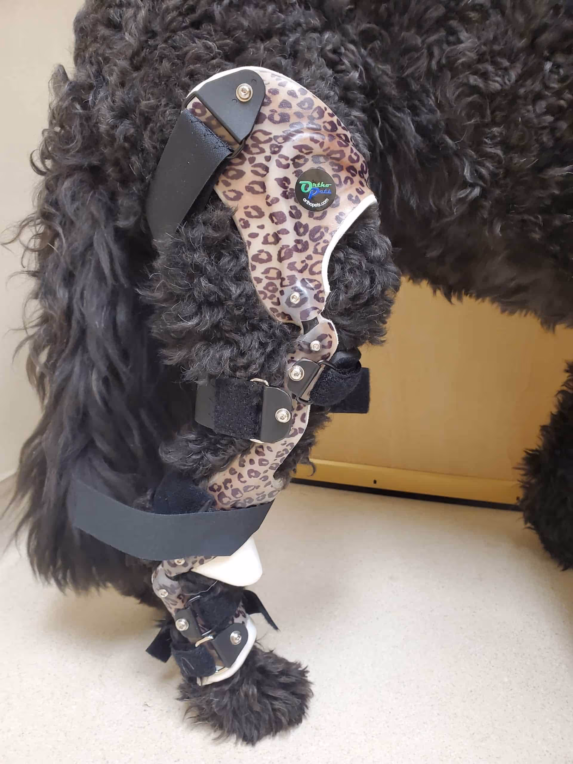
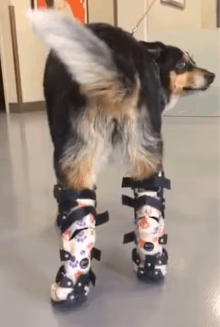
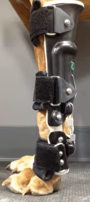
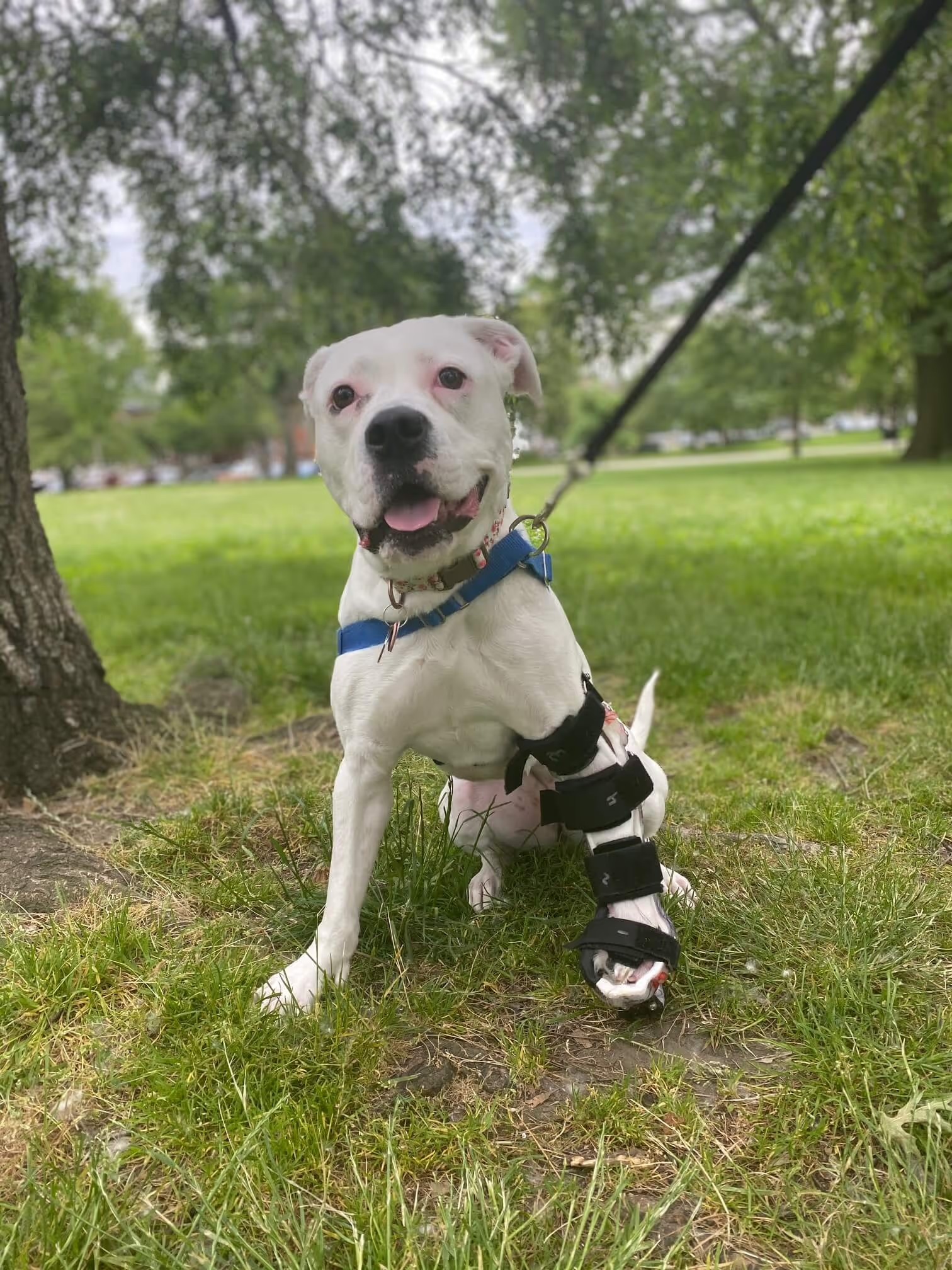
Fig. 6. An example of canine carpal orthosis. This device is used for mild to
moderate carpal hyperextension or collateral ligament injury (carpal varus or
valgus).[/caption]An example is shown in Fig 6. This device is designed to limit carpal extension while allowing flexion; it is used for carpal hyperextension owing to palmar fibrocartilage damage or injury to the flexors of the carpus (muscles or tendon), or both. At full extension (101), the device locks, resisting further carpal exten- sion. This can be thought of as an arthrodesis on demand. The advantages over an arthrodesis are (1) functional range of motion of the digits, (2) range of motion of the carpus within safe limits, (3) removable for rehabilitation and skin management, and (4) dynamism allowing initial complete restriction of carpal flexion followed by sequential return of range of motion. This design can be used for frontal plane instability as well. In this case, the shell resists varus and valgus while full carpal range of motion in the sagittal plane is allowed by virtue of the polycentric hinge.Importantly, with significant instability, soft wraps made of fabric, neoprene, bandage material, etc., do not have enough structural rigidity to resist joint collapse. Significant instability means more than 51-101 difference in range of motion relative to the contralateral limb. As such, these products are reserved forcompression, proprioceptive cuing (so-called Kinesio Taping), and warmth.